Not-so-restful resting connectivity fMRI (pt.1)
Last week, during a very interesting Brain, Behavior and Cognition colloquium given by Steve Nelson, Jeff Zacks asked a thought-provoking question. He wanted to know what fMRI connectivity maps would look like if you performed a resting-connectivity-type analysis on data that wasn't collected at rest, but was instead collected whilst participants were 'on-task', i.e. doing things other than resting.
As background to this:
- Each fMRI participant in our lab typically carry out one or two connectivity scans prior to, or following a the bulk of the experimental scanning;
- Connectivity scans require that the participant keep their eyes open, fixate on a fixation cross, and try not to fall asleep for about 5 minutes;
- Experimental scans have participants engage in much more effortful cognitive processes (such as trying to recognise words and scenes), with multiple tasks presented in relatively quick succession;
- Resting connectivity is thought to be driven be low frequency fluctuations in signal ( approx. 0.1 Hz; a peak every 10 seconds or so), on-task BOLD activation is much more event-related, ramping up or down following certain events which occur every 6 seconds or so (which we assume results from the engagement of a particular cognitive process).
- This Raichle article in Scientific American is a a very accessible primer on the current state of resting connectivity. This Raichle article in PNAS is a more comprehensive scientific discussion of the same topic.
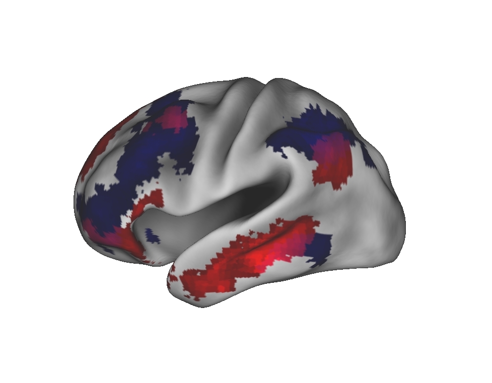
Jeff's question was interesting to me because it asks how robust these slow-wave oscillations across distal brain loci really are. To what extent would they be (un-)modulated by task-related fluctuations in BOLD signal?
My initial thoughts were that on-task resting connectivity maps would look pretty similar to resting resting connectivity maps - after all, it has been suggested that resting connectivity networks, such as the fronto-parietal control network, arise because of their frequency of coactivation during development, i.e. that DLPFC, MPFC and IPL are coactive when on-task so often that it makes metabolic sense for their activity to synchronise even when not on-task. But, there's no need to be satisfied with your initial thoughts when you can simply look at some data, so that's what I did.
On Friday I began the process of running a resting state-style connectivity analysis on the on-task scans of the data that went into the Journal of Neuroscience paper we had published a few weeks ago. It was a nice dataset to use as we had also collected resting scans and carried out a connectivity analysis that yielded some interesting results. I entered the same two seeds (from the the PFC mid-line) that were central to our connectivity analysis into a connectivity analysis using the four, 10-minute on-task scans that we analysed for the event-related fMRI analysis. In part two, I'll have an informal look at the differences between the output from the resting scans and the on-task scans when subjected to the same resting connectivity analyses.
Originally published 5 April 2010 at akiraoconnor.org/2010/04/05/not-so-restful-resting-connectivity-fmri-pt-1/